Research/Theme 04

Neuroinflammation and glial responses

We develop and apply imaging approaches to study neuroinflammation and glial responses in models of neurological and psychiatric disease, including the molecular targets TSPO and CSF1R.

Tracer developmentExperimental modelsPETAutoradiographyHistology
Schematic The tracer-development programme, as a pipeline Three stations linked by arrows. First, candidate tracers, drawn as a vial of generic tracer molecules. Second, experimental models, drawn as a brain section with a small local lesion. Third, imaging and tissue validation, drawn as a PET scanner ring, a glass slide with a brain section, and a glial cell. A dashed arrow runs from the last station back to the first: what the signal means feeds back into choosing tracers. Generic drawing; no tracer structures and no data. WHAT THE SIGNALMEANS FEEDS BACK 1 CANDIDATE TRACERS AND NEW TARGETS 2 EXPERIMENTAL MODELS E.G. A LOCAL LESION 3 IMAGING + TISSUE VALIDATION PET · AUTORADIOGRAPHY · HISTOLOGY

At a glance

Most of our current work in this theme is not yet published, so this page describes the questions we ask and how we test them. Findings are added here once they are published.

Main biological question
How can we develop and validate imaging tools that better characterise the brain's inflammatory and glial responses?
What we are investigating now
Developing and validating new tracers for neuroinflammation and glial responses, including targets beyond TSPO such as CSF1R; experimental models, including quinolinic acid lesions; and astrocyte-associated imaging, for example with [3H]BU99008, which binds imidazoline-2 sites and is used to investigate astrocyte-associated changes, with tissue validation. We also ask whether changes in GPR6 signalling are associated with neuroinflammatory responses (below).
Approaches and models
Autoradiography and blocking studies on brain sections, then PET; histology (Iba1, MHCII or HLA-DR, GFAP) and inflammatory messengers in brain, blood and cerebrospinal fluid. Rats, mice, minipigs and post-mortem human tissue.
Established in published work
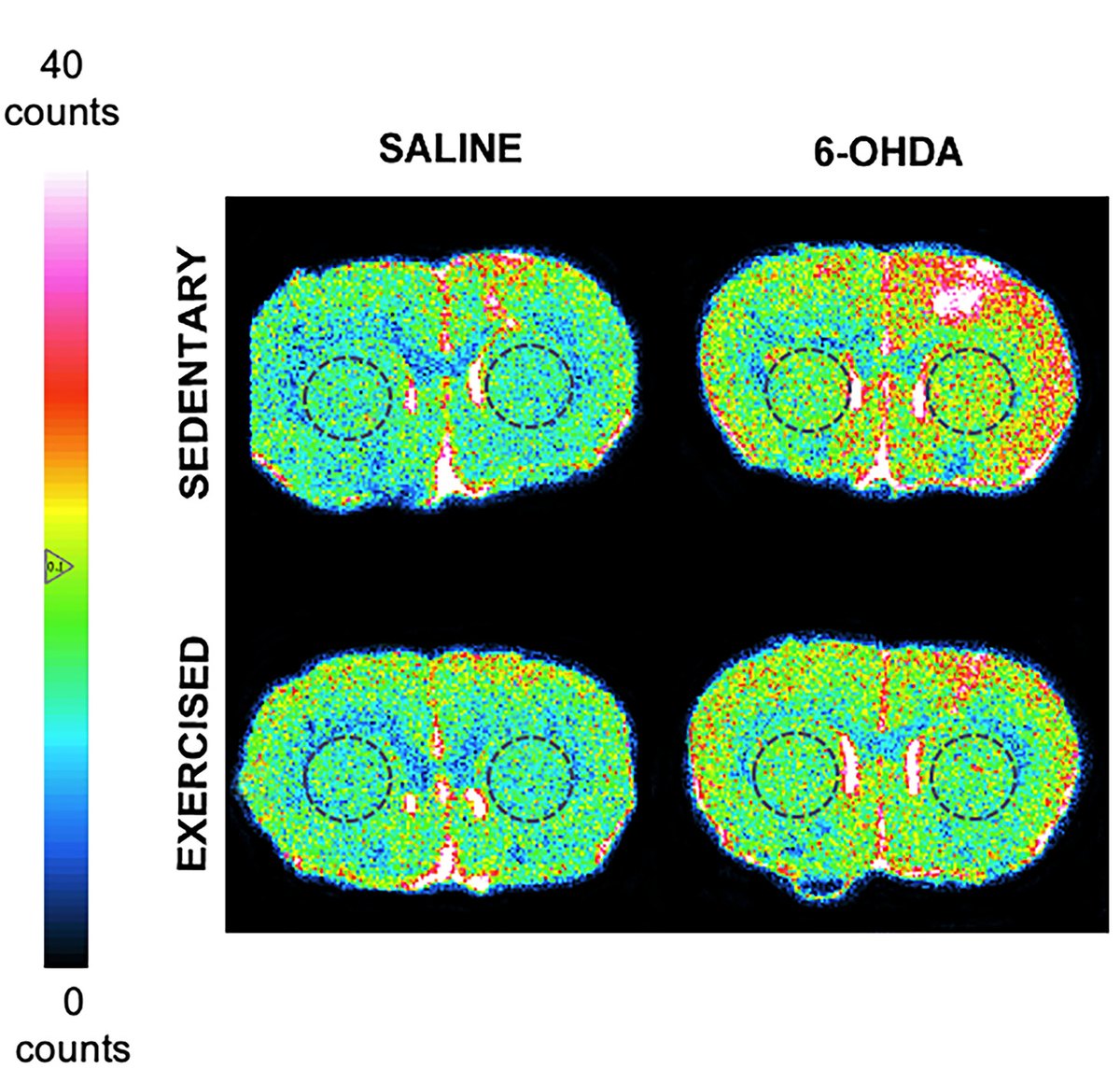
In published studies, TSPO binding was higher in α-synuclein models in rats and minipigs, in sedentary but not exercised 6-OHDA-lesioned rats, in Alzheimer's frontal cortex and in a rat model of fatty liver disease; how TSPO binding is interpreted depends on species, model and cellular context. A collaborative study characterised [3H]5-MOP as a selective CSF1R radioligand (Iavazzo et al. 2026).
Who is involved

Elisa Mazzoleni and Miriam Fiorino use [³H]PK11195 autoradiography to measure TSPO binding within the GPR6 project.

Useful collaborations
Tracer chemistry for new glial targets; models with well-characterised glial responses; tissue that can be paired with histology. How to start a project →

01 The question

How can we develop and validate imaging tools that better characterise the brain's inflammatory and glial responses?

Read moreShow less: How can we develop and validate imaging tools that better characterise the brain's inflammatory and glial responses?

Glial cells, including microglia, the brain's resident immune cells, and astrocytes, respond to injury, to protein aggregates such as α-synuclein, and to inflammation elsewhere in the body. This response, neuroinflammation, accompanies Parkinson's disease, Alzheimer's disease and other brain disorders.

The most widely used imaging target for it has been the translocator protein TSPO, which sits on mitochondria. TSPO binding often rises where glial cells respond, but it is not a direct or universal measure of microglial activation: which cells carry the signal, and what a change means, depend on the species, the disease model and the cellular context. That is why we check it against tissue staining, and why new tracers and targets are being sought.

Our published TSPO studies are an important foundation. Our current focus is on developing and validating new tracers for neuroinflammation and glial responses, including targets beyond TSPO, such as CSF1R.

We combine imaging and autoradiography with molecular and histological analyses to understand the cellular processes underlying these signals and their responses to disease and treatment.
02 Current research

What we are investigating, and how

Read moreShow less: What we are investigating, and how

The programme runs along three connected lines, following the pipeline at the top of the page: candidate tracers, the models they are tested in, and the measurements that show what their signal means.

1TracersOngoing

Tracer development and validation

Evaluating new candidate tracers and complementary molecular targets to improve how we measure and interpret neuroinflammation.

  • TargetsTSPO, the established target, and targets beyond it, such as CSF1R, a receptor found on microglia.
  • ApproachTest where a candidate binds and whether the binding is specific, with autoradiography and blocking studies on brain sections; then whether it reaches the brain and gives a useful measurement with PET.
2ModelsOngoing

Experimental models

Including our quinolinic acid (QA) lesion work, which provides a setting for investigating tracer binding alongside tissue injury and glial responses.

  • A model we knowQuinolinic acid makes a local lesion. We have used QA lesions in published SV2A imaging, comparing PET and autoradiography on the lesioned and intact sides.
  • Why it helpsA local lesion puts tracer binding, tissue injury and the glial response side by side in the same brain.
QA lesions in Theme 02 →
3SignalOngoing

Understanding the signal

Combining PET and autoradiography with histology and molecular analyses to investigate what changes in binding actually represent.

  • LevelsThe living brain, brain sections, the cells themselves, and the messengers they release.
How the levels fit together ↓
Under investigation

Astrocyte-related imaging

Alongside microglia, we are investigating imaging approaches for astrocytes.

Our autoradiography toolbox includes [3H]BU99008, which binds imidazoline-2 sites and is used to investigate astrocyte-associated changes. What a change in its binding means for astrocytes has to be validated in tissue, for example with astrocyte staining. See the toolbox →

03 Understanding the signal

What does a change in binding actually represent?

Read moreShow less: What does a change in binding actually represent?

A change in tracer binding tells us that something has changed, not which cells changed or what they are doing.So each signal is checked against the cells themselves, in tissue from the same animals.

  1. PETLiving brain

    Binding in the whole brain, over time

    Follows a tracer in the same animal before and after a change. We have used [11C]PK11195 PET in minipig models of Parkinson's disease.

    Lillethorup et al., Synapse 2018 · doi
  2. AutoradiographyBrain sections

    Where, and how specific

    Maps binding at far finer detail. Neighbouring sections incubated with an excess of an unlabelled drug show how much of the binding is specific.

    Binda et al. 2021 · Stokholm et al. 2021 · Kjærgaard et al. 2024 · Metaxas et al. 2019
  3. HistologyThe cells

    Staining the glia

    Antibodies show the cells behind a signal: Iba1, MHCII or HLA-DR for microglia, GFAP for astrocytes.

    Kjærgaard et al. 2024 · Thomsen et al. 2021 · Lillethorup et al. 2018
  4. MolecularSignals

    Inflammatory messengers

    Cytokines and chemokines measured in brain, blood and cerebrospinal fluid.

    Kjærgaard et al. 2024 · Metaxas et al. 2019

The examples are from our published TSPO work. The same levels are how we examine new tracers.

04 Selected published work

The foundation: what our published studies show

Read moreShow less: The foundation: what our published studies show

Our published studies used TSPO to follow glial responses in models of Parkinson's disease, in Alzheimer's disease tissue and in a model of fatty liver disease. Start with one study, section by section.

Read the sections · a lesion, with and without exerciseDemo playing · press to take over
Four colour-coded autoradiograms of coronal rat brain sections through the striatum, arranged by injection (saline, 6-OHDA) and group (sedentary, exercised), beside a colour scale from 0 to 40 counts. The sedentary 6-OHDA section shows higher binding, in red, over one hemisphere. Saline · sedentary

[3H]PK11195 autoradiography of TSPO binding in coronal rat brain sections through the striatum; representative sections, colour scale 0–40 counts; dashed circles mark the striatum. Rats were injected in one striatum with saline or 6-OHDA, then either stayed sedentary or ran on a treadmill for five weeks. Binda KH, Lillethorup TP, Real CC, Bærentzen SL, Nielsen MN, Orlowski D, Brooks DJ, Chacur M, Landau AM. Exp Neurol 2021;342:113741 · Figure 3A · doi:10.1016/j.expneurol.2021.113741 · CC BY 4.0; cropped, panel letter removed; highlight and shading added by us.

Glial responses in our published studies

Across species and models, a glial response often appeared early, sometimes before other damage could be measured.

Parkinson's disease models

  1. ↑

    α-Synuclein in minipigs. After a gene for mutant human α-synuclein was delivered to the substantia nigra, [11C]PK11195 binding was higher in the basal ganglia and cortical areas than in control minipigs, with no change in dopamine markers and no motor symptoms. The authors suggest microglial activation is an early response to α-synuclein.

    PETMinipigLillethorup et al., Synapse 2018 · doi

  2. ↑

    α-Synuclein in rats. With human α-synuclein overexpressed, [3H]PK11195 binding on the injected side rose 9.8% in the striatum and 13.9% in the substantia nigra, significantly more than in control rats given a GFP vector.

    AutoradiographyRatStokholm et al., Biomedicines 2021 · doi

  3. ↑

    α-Synuclein fibrils in rats. After fibrils were injected into one striatum, MHCII-positive microglia appeared early and persisted in the striatum and substantia nigra on the injected side.

    HistologyRatThomsen et al., Neurobiol Dis 2021 · doi

  4. ↑

    6-OHDA, with and without exercise. TSPO binding rose in the injected striatum of sedentary rats. In lesioned rats that exercised on a treadmill, the rise was not present.

    AutoradiographyRatBinda et al., Exp Neurol 2021 · doi

  5. ↑

    Proteasome inhibition in minipigs. After lactacystin was injected into the medial forebrain bundle, activated (HLA-DR-positive) microglia appeared in the substantia nigra and nearby areas on the injected side.

    HistologyMinipigLillethorup et al., Exp Neurol 2018 · doi

Alzheimer's disease

  1. ↑

    Post-mortem frontal cortex. [3H]PK11195 binding was higher in the grey matter of patients than of non-demented subjects, and went up with tau pathology. SV2A binding did not differ. Theme 02 →

    AutoradiographyHuman tissueMetaxas et al., Front Cell Neurosci 2019 · doi

  2. ↑

    Amyloid mice. In ageing APPswe/PS1dE9 mice, raised [3H]PK11195 binding and IL-1β came before the loss of serotonin transporter density and activity in the neocortex.

    Autoradiography · molecularMouseMetaxas et al., Alzheimers Res Ther 2019 · doi

Body and brain

  1. ↑

    Fatty liver disease. Rats fed a high-fat, high-cholesterol diet developed fatty liver disease and inflammation in the blood. In prefrontal cortex, [3H]PK11195 binding was 19% higher, matching microglial changes seen with Iba1 staining, and SV2A binding was lower. The rats also showed impaired memory and depression-like behaviour.

    Autoradiography · histologyRatKjærgaard et al., JHEP Rep 2024 · doi

Published figure with two panels. A: [3H]PK11195 autoradiograms of rat brain sections at the striatum, GFP control and α-synuclein, with a graph showing binding on the injected side as a percentage of the other side: minus 1.94% for GFP, 9.80% for α-synuclein. B: the same at the substantia nigra: minus 1.09% for GFP and 13.9% for α-synuclein.
TSPO binding after α-synuclein overexpression. [3H]PK11195 autoradiograms and binding on the injected side as a percentage of the other side, at the level of the striatum (A) and substantia nigra (B), in rats given α-synuclein (ASYN) or a control GFP vector. Stokholm K, Thomsen MB, Phan JA, Møller LK, Bay-Richter C, Christiansen SH, Woldbye DPD, Romero-Ramos M, Landau AM. Biomedicines 2021;9(12):1876 · Figure 7 · doi:10.3390/biomedicines9121876 · CC BY 4.0; resized.

Beyond TSPO: a tracer for CSF1R

Published · EJNMMI Research 2026

In a collaborative study, [3H]5-MOP was characterised as a radioligand for CSF1R. In autoradiography of human meningioma tissue it bound a single saturable site with high affinity (KD 9.8 nM), and structurally different CSF1R inhibitors displaced it. The authors report that 5-MOP was selective for CSF1R across a large panel of kinases. In mouse brain five days after an ischaemic stroke, [3H]5-MOP binding was confined to the lesion.

Iavazzo C, et al. [3H]5-MOP: a novel and selective colony stimulating factor-1 receptor (CSF1R) radiotracer. EJNMMI Research 2026 · doi:10.1186/s13550-026-01520-x

Papers in this theme

  1. 2026
  2. 2024
  3. 2021
    Binda KH, Lillethorup TP, Real CC, … Chacur M, Landau AM · Experimental Neurology
  4. 2021
    Stokholm K, Thomsen MB, Phan JA, … Romero-Ramos M, Landau AM · Biomedicines
  5. 2021
    Thomsen MB, Ferreira SA, Schacht AC, … Landau AM, Romero-Ramos M · Neurobiology of Disease
  6. 2019
    Metaxas A, Thygesen C, Briting SRR, Landau AM, Darvesh S, Finsen B · Frontiers in Cellular Neuroscience
  7. 2018
    Lillethorup TP, Glud AN, Landeck N, … Landau AM · Synapse

All neuroinflammation papers →Tritium tracer toolbox →

Questions for review

  1. Can CSF1R now be named publicly, as in the lede and the Current research section?
  2. Is it OK to name the quinolinic acid (QA) lesion work as a model we are using?
  3. Is "astrocyte-related imaging … under investigation" worded right, and can [3H]BU99008 stay?
  4. [3H]5-MOP is now cited as the published paper (Annie, 9 Oct 2026) and is in the publication archive once. The stroke-model sentence is in the published article (Results, Fig. 6), so it stays.
  5. Does the pipeline drawing (candidate tracers → experimental models → imaging and tissue validation) describe the programme well?