Draft for review Prototype prepared for the Landau Lab · September 2026 · not yet final

Methods/Imaging methods and biological validation

From signal to evidence

No single measurement tells the whole story. We combine PET and quantitative autoradiography with behavioural testing and analyses of brain tissue — so that each can test, sharpen or explain what the others show.

PETAutoradiographyTissue analysesBehaviourRodents & minipigs
Three overlapping circles — in vivo PET, autoradiography and tissue analyses, and behaviour. Where they overlap is where evidence is strongest. IN VIVO PET THE LIVING BRAIN AUTORADIOGRAPHY & TISSUE BEHAVIOUR FUNCTION evidence

01 Our approach

Understanding what imaging signals represent

We investigate how brain disorders and treatments affect molecular signalling and brain function, with a particular focus on Parkinson's disease and depression.

Using positron emission tomography (PET) and quantitative autoradiography in rodents and minipigs, we study molecular targets in the living brain and in brain tissue. We combine imaging with behavioural testing and neurochemical, molecular and histological analyses to understand what imaging signals represent biologically and how they change with disease and treatment.

These methods are not steps in a fixed sequence. Which one comes first depends on the question — and a study often returns to an earlier method once another has raised a new question.

  • Start in tissue

    Map where a tracer binds, and check that the binding is specific, at high resolution on brain sections — then take it into the living brain. This is how we are approaching GPR6. GPR6 →

  • Start in the living brain

    Follow a change over time with PET in the same animal, then ask in tissue what it reflects. In rat lesion models, [11C]UCB-J PET and [3H]UCB-J autoradiography found closely matching losses of SV2A binding.

  • Start with function

    Begin with an intervention and a behavioural read-out, then look for its molecular correlates. In a rat Parkinson's model, treadmill exercise was followed by pain-threshold testing and autoradiography of four targets.

02 Three kinds of measurement

What each method sees — and what it can't

[11C]yohimbine PET parametric maps of a minipig brain in three views, at baseline, 8 minutes and 4 hours after nisoxetine; colour scale 2.0 to 6.0 mL/ccm.PET

In vivo · the living brain

PET

Measures
Where a radiolabelled tracer goes in the living brain, over time. With kinetic modelling, that becomes a quantitative measure of binding to a target.
Strength
Non-invasive and repeatable in the same animal — before and after treatment, or as disease develops. The same method is used in people.
Alone, can't
Resolve fine anatomical detail, or say which cellular change lies behind a change in binding.

[11C]yohimbine VT maps in a minipig at baseline and after nisoxetine, a drug that raises noradrenaline. Landau et al., Biomolecules 2023 · doi:10.3390/biom13040674 · CC BY 4.0, cropped.

Four coronal rat brain autoradiograms of [3H]UCB-J binding at striatal level, arranged saline versus 6-OHDA and sedentary versus exercised, with dashed circles over the left and right striatum.Autoradiography

Ex vivo · brain sections

Autoradiography & tissue analyses

Measures
Radioligand binding on thin brain sections, quantified against standards — together with histological, molecular and neurochemical analyses of the same brains.
Strength
Far finer spatial detail than PET. Specificity can be checked directly by blocking, and several targets can be measured in the same brains.
Alone, can't
Follow change over time: each brain gives one time point, and binding is measured under conditions that differ from the living brain.

[3H]UCB-J autoradiograms (SV2A) in saline- and 6-OHDA-lesioned rats, sedentary or exercised; warmer colours mean more binding (relative scale). Binda et al., Exp Neurol 2021 · doi:10.1016/j.expneurol.2021.113741 · CC BY 4.0, cropped.

Photo to come

Behavioural testing in the lab

Function · what animals do

Behaviour

Measures
What animals do — for example motor function, sensitivity to pain, or behaviours used to model aspects of depression.
Strength
Connects molecular findings to function, and to the symptoms that treatments aim to change.
Alone, can't
Explain mechanism. Behaviour has many causes, and animal models capture selected features of a human disorder, not the whole condition.

We also use neurochemical measures in living animals — for example microdialysis — and MRI through collaborations.

03 How they fit together

Start from the question, not the method

Choose a question to see which measurements answer it, how they check one another, and an example from our published work.

    PETLIVING BRAIN TissueAUTORADIOGRAPHY& ANALYSES BehaviourFUNCTION OTHERMEASURES No arrows: any measurement can come first.

    04 Rodents and minipigs

    Choosing the model for the question

    Rodents

    Rats and mice let us combine imaging with detailed behavioural testing and tissue analyses in larger groups, and use genetic and pharmacological approaches to change a target directly.

    Small-animal PET makes it possible to scan the same rat repeatedly — for example before and after a treatment — while autoradiography of the same models maps binding at high resolution.

    Göttingen minipigs

    Minipigs have larger, folded brains that can be scanned on clinical PET/CT systems, with arterial blood sampling for full quantification. In our [11C]UCB-J study, minipig VT values were closer to reported human values than those derived from rhesus monkeys.

    Large-animal imaging raises its own methodological questions, and we have studied several of them directly:

    • How the choice of anaesthetic influences tracer binding
    • Automated arterial blood sampling for PET, and its risks for the animals
    • Quantitative gait analysis to measure motor function
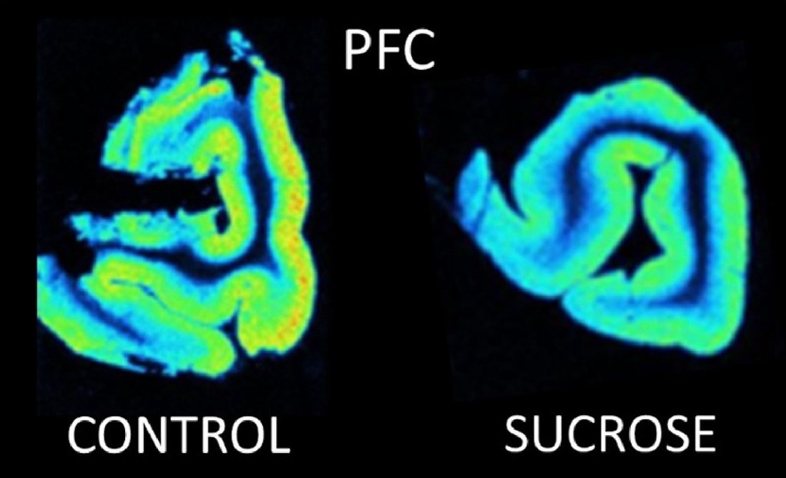
    Two pseudo-colour autoradiograms of minipig prefrontal cortex, labelled control and sucrose; the folded cortical ribbon shows more green and yellow in the control section and more blue in the sucrose section.
    [3H]UCB-J autoradiography of minipig prefrontal cortex. After 12 days of daily sucrose, SV2A binding in this region was lower than in controls. The folded cortex of the minipig brain is visible in both sections; no calibrated colour scale is shown. Bærentzen SL, Thomsen MB, Alstrup AKO, Wegener G, Brooks DJ, Winterdahl M, Landau AM. Neuropharmacology 2024;256:110018 · doi:10.1016/j.neuropharm.2024.110018 · CC BY 4.0, cropped.

    05 Collaborations

    An integrated platform

    Our platform brings together PET, quantitative autoradiography, behavioural testing and analyses of brain tissue. Collaborations in radiochemistry, MRI, molecular biology, histology and neurosurgery support tracer development and validation, experimental interventions and interpretation of imaging findings.

    RadiochemistryMRIMolecular biologyHistologyNeurosurgery

    This integrated approach connects molecular measurements with brain function, behaviour and treatment effects.

    06 Methods papers

    Selected methods and validation work

    1. 2023
      Landau AM, Jakobsen S, Thomsen MB, … Doudet DJ · Biomolecules
    2. 2021
      Thomsen MB, Jacobsen J, Lillethorup TP, … Brooks DJ, Landau AM · Journal of Cerebral Blood Flow & Metabolism
    3. 2020
      Thomsen MB, Schacht AC, Alstrup AKO, … Brooks DJ, Landau AM · Molecular Imaging and Biology
    4. 2021
      Binda K, Lillethorup T, Real C, … Chacur M, Landau A · Experimental Neurology
    5. 2022
      Nielsen SV, Dollerup MR, Bærentzen SL, Landau AM, Munk OL, Alstrup AKO · Laboratory Animals
    6. 2022
      Steinmüller JB, Binda KH, Lillethorup TP, … Sørensen JCH, Glud AN · Journal of Neuroscience Methods
    7. 2020
      Landau AM, Noer O, Alstrup AKO, … Doudet DJ, Winterdahl M · Molecular Imaging and Biology
    8. 2013
      Alstrup AKO, Landau AM, Holden JE, … Doudet DJ · BioMed Research International
    9. 2023
      Real CC, Binda KH, Thomsen MB, Lillethorup TP, Brooks DJ, Landau AM · Current Neuropharmacology

    All publications: publication list →

    Questions for review

    1. Are these the right example studies to feature, or would you prefer others?
    2. Which equipment and facilities should the page name — and which belong to shared facilities or collaborators?
    3. Do you have photos of behavioural testing, the scanners or the lab that we could use?