Methods/Models
Rodents and minipigs
Rats, mice and Göttingen minipigs, each chosen for what the question needs.
01 Rodents and minipigs
Choosing the model for the question
Rodents
Rats and mice let us combine imaging with detailed behavioural testing and tissue analyses in larger groups, and use genetic and pharmacological approaches to change a target directly.
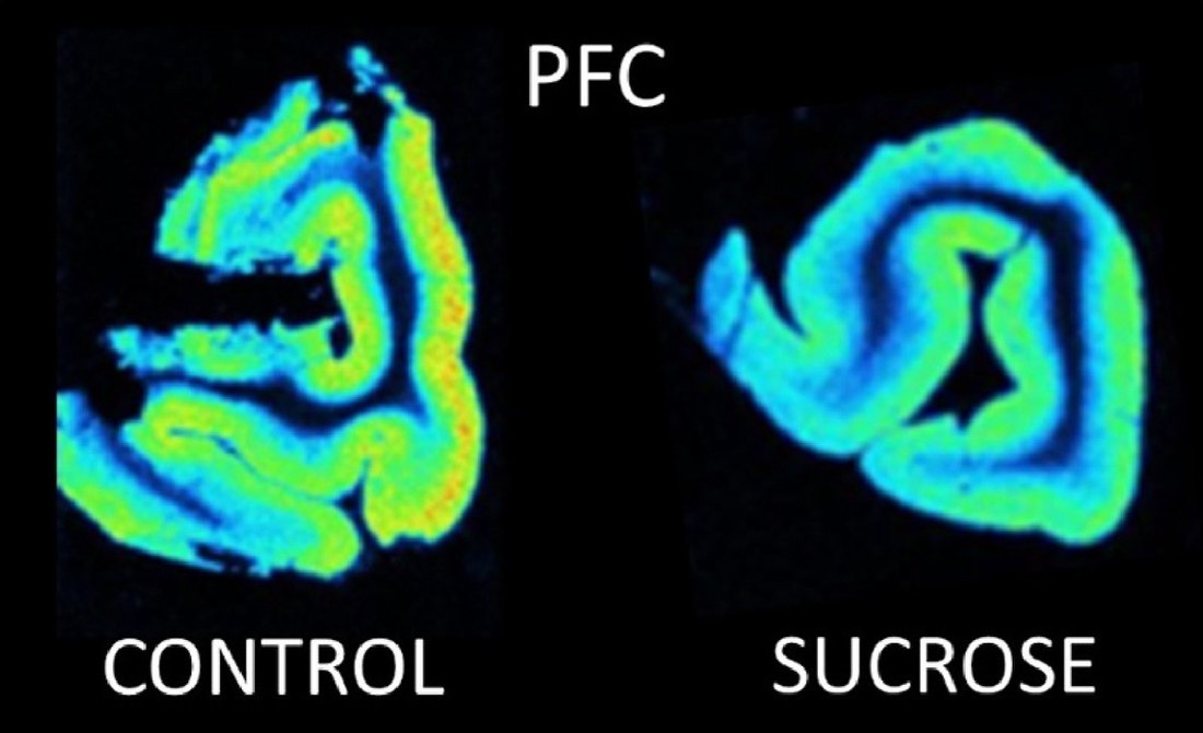
Small-animal PET makes it possible to scan the same rat repeatedly — for example before and after a treatment — while autoradiography of the same models maps binding at high resolution.
Göttingen minipigs
Minipigs have larger, folded brains that can be scanned on clinical PET/CT systems, with arterial blood sampling for full quantification. In our [11C]UCB-J study, minipig VT values were closer to reported human values than those derived from rhesus monkeys.
Large-animal imaging raises its own methodological questions, and we have studied several of them directly:
- How the choice of anaesthetic influences tracer binding
- Automated arterial blood sampling for PET, and its risks for the animals
- Quantitative gait analysis to measure motor function